


When you first hear the words “keratoconus,” it can be a lot to take in. The good news is that modern treatments are incredibly effective at protecting your sight. The key is to understand that we manage this condition with a clear, two-part strategy: first, we stop the cornea from getting weaker, and second, we work on getting your vision as clear as possible. This dual approach is the bedrock of every successful treatment plan.
Understanding Keratoconus and Your Treatment Goals
Think of your eye as a sophisticated camera and the cornea as its primary lens. In a healthy eye, that lens has a perfect dome shape, focusing light precisely to give you a sharp picture. With keratoconus, the cornea thins and starts to bulge outward, forming a cone. Just like a warped camera lens would create a distorted photo, an irregular cornea blurs and warps what you see.
Getting this diagnosis naturally brings up a lot of questions. Keratoconus is a progressive condition, meaning it can worsen over time if it’s not managed. Recent research underscores its growing prevalence, which has climbed to roughly 1,155.2 cases per 100,000 people globally since 2020. It most often affects young adults between 20 and 29, which is why catching it early and being proactive is so important. You can find more details on this trend in the Review of Optometry.
The Two Pillars of Treatment
To manage keratoconus effectively, your eye care team will concentrate on two separate but equally vital goals. This framework makes it easier to understand why certain treatments are recommended at specific points in your care.
- Goal 1: Halt Progression. Our absolute first priority is to stabilize the cornea. We need to stop the cone shape from getting any worse. Think of it like reinforcing the foundation of a building to prevent it from shifting.
- Goal 2: Improve Vision. Once the cornea is stable, we shift our focus to correcting the blurry and distorted vision its irregular shape has caused. This step is all about creating a new, smooth surface for light to travel through cleanly.
“A new diagnosis of keratoconus is a lifelong journey. The most critical message for patients is that we have a two-part treatment plan: we first stabilize the eye with cross-linking, then we improve vision with specialty contact lenses.”
This structured approach makes sure we’re addressing both the underlying structural issue and the vision problems it creates. Your first and most crucial step is finding a specialist who can guide you through this process. When you start your search, you can learn more about finding a keratoconus doctor near you.
The table below breaks down these goals and the treatments that help us achieve them.
Keratoconus Treatment Goals at a Glance
This table outlines the primary objectives of keratoconus management and the corresponding treatment categories designed to achieve them.
| Treatment Goal | Primary Approach | Common Therapies |
|---|---|---|
| Halt Progression | Strengthen the cornea to stop the bulging process. | Corneal Cross-Linking (CXL) |
| Improve Vision | Create a smooth optical surface for clear focus. | Specialty Contact Lenses (RGP, Scleral), Intacs, Corneal Transplant |
By breaking the journey down into these two clear goals, managing keratoconus becomes a much more straightforward and less intimidating process.

“Most of my keratoconus patients get their best vision from specialty contact lenses. Regular glasses often can’t correct an irregular cornea well enough.”
Improving Vision with Non-Surgical Solutions
Once we’ve successfully halted the progression of keratoconus, our focus shifts to the next critical goal: restoring clear vision. The irregular, cone-like shape of the cornea in keratoconus severely distorts the light entering your eye. This is why standard glasses and off-the-shelf soft contact lenses just don’t cut it anymore; they can’t compensate for the unique, bumpy surface the condition creates.
The strategy here is to create a brand-new, perfectly smooth optical surface in front of the cornea. A good analogy is paving a bumpy, uneven road with a fresh, flawless layer of asphalt. Suddenly, the ride is perfectly smooth. In eye care, our “asphalt” is a specialty contact lens, which is the key to delivering the crisp, clear vision that glasses simply can no longer provide.
This simple infographic breaks down the two main goals we tackle when treating keratoconus.
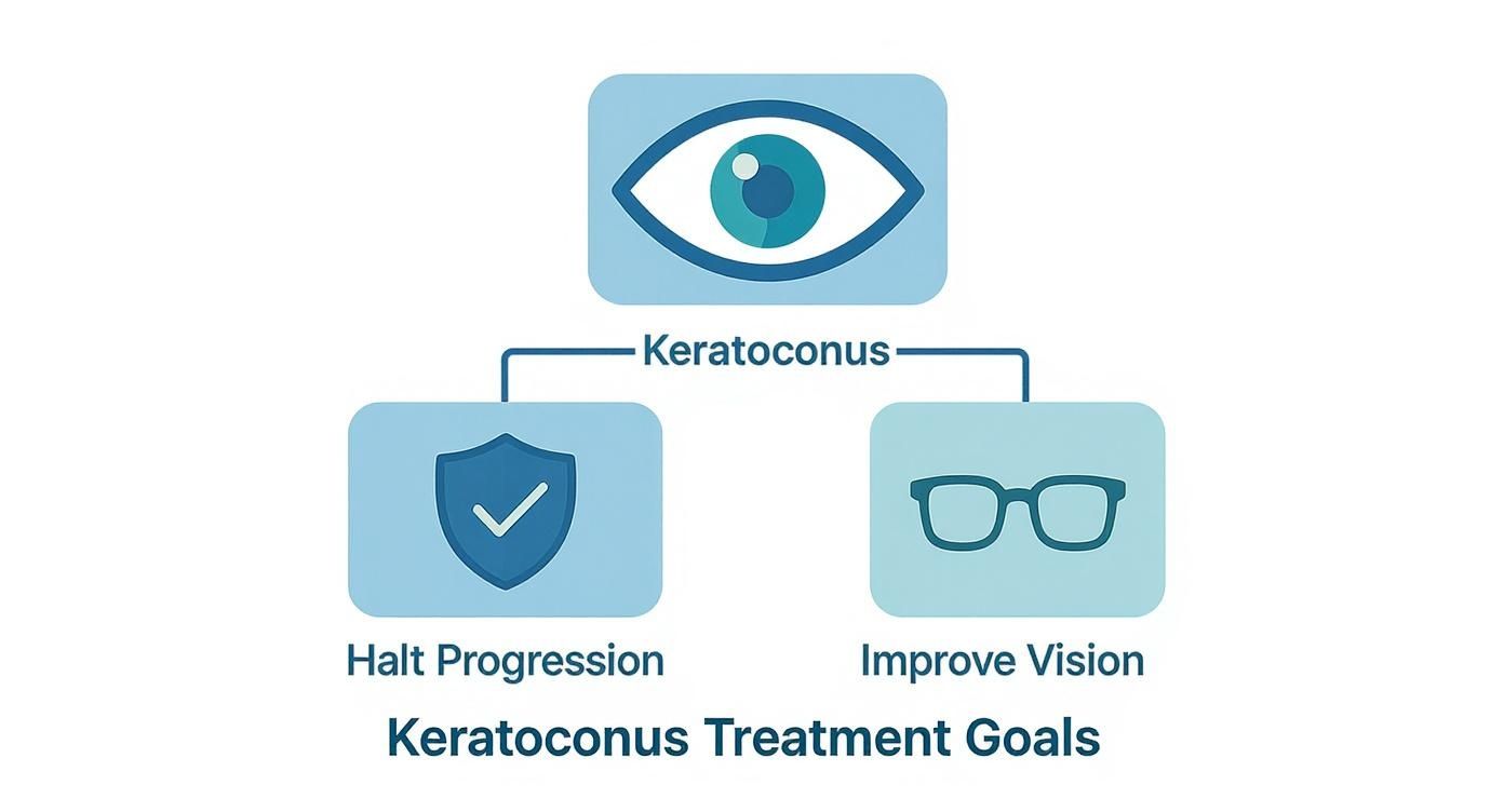
As you can see, halting progression and improving vision are two distinct but parallel paths. This section is all about that second goal—getting you to see clearly again.
Rigid Gas Permeable Lenses
Rigid Gas Permeable (RGP) lenses are often one of the first specialty options we turn to. These are made from a firm, breathable material that holds its shape on the eye, rather than draping over the cornea’s irregular surface like a soft lens would.
This rigidity is exactly what makes them so effective. An RGP lens vaults over the cone-shaped part of the cornea, and the tiny space between the back of the lens and the front of your eye fills with natural tears. This tear layer neutralizes the corneal irregularities, creating that new, smooth surface we need for sharp vision.
The trade-off? Some patients find RGPs take a little getting used to. Because they are smaller and move slightly with each blink, you might be more aware of the lens edge, especially at first.
Hybrid Lenses: The Best of Both Worlds
For anyone who needs the crystal-clear optics of an RGP but struggles with the initial comfort, hybrid lenses are a fantastic solution. The name says it all—they are a hybrid of two distinct materials.
The center of the lens is made from a crisp, rigid gas permeable material for that unbeatable sharpness. This is fused directly to a soft, flexible outer skirt, much like a regular soft contact lens. This design gives you the comfort of a soft lens with the superior visual quality of a hard one. The soft skirt also helps center the lens and keep it stable, which cuts down on the movement and edge awareness people sometimes feel with RGPs.
Hybrid lenses beautifully bridge the gap between comfort and clarity. By combining a rigid center with a soft outer ring, they deliver sharp, stable vision without sacrificing the comfortable wearing experience many patients desire.
This unique combination makes them an excellent choice for people with active lifestyles or sensitive eyes who need a reliable, comfortable way to manage their keratoconus.
Scleral Lenses: The Ultimate Liquid Shield
Perhaps the most exciting and effective tool in our non-surgical arsenal is the scleral lens. These are large-diameter lenses that do something remarkable: they don’t touch the sensitive cornea at all. Instead, they vault completely over it, resting gently on the sclera (the white part of your eye).
Think of a scleral lens as a personal “liquid shield” for your eye. Before you put it in, the bowl of the lens is filled with a sterile, preservative-free saline solution. Once it’s on your eye, this saline creates a constant reservoir of fluid that bathes the cornea all day long.
This fluid-filled chamber offers two incredible benefits:
- Perfect Optics: The liquid fills in every single irregularity on the corneal surface, creating a perfectly smooth and uniform window for light to pass through. This often produces vision that is sharper and clearer than what can be achieved with any other method.
- Therapeutic Comfort: For patients who also battle dry eye—a very common issue alongside keratoconus—this constant hydration provides profound relief and comfort that lasts all day.
Because they are large and exceptionally stable, most people find scleral lenses to be the most comfortable option available. Your eyelids glide smoothly over the surface without ever interacting with a lens edge. Their size also provides a bonus shield against environmental irritants like dust and wind.
For a deeper look into how these advanced lenses work, feel free to explore our detailed guide on contact lenses in keratoconus. Ultimately, choosing the right lens is a highly collaborative process between you and your eye doctor, tailored to your unique corneal shape, the stage of your condition, and your day-to-day lifestyle.
Stopping Progression with Corneal Cross-Linking
While specialty contact lenses are incredible tools for improving vision, they don’t solve the underlying problem: the progressive weakening of the cornea. To get ahead of keratoconus, we first have to stop it in its tracks. This is where a procedure called corneal cross-linking (CXL) makes a world of difference. It’s currently the only treatment proven to halt the progression of the disease and prevent further vision loss.

Think of your cornea as a piece of fabric held together by countless tiny collagen threads. In keratoconus, those threads weaken, causing the fabric to stretch and bulge. CXL works by reinforcing this very fabric, making it strong and stable once more.
The procedure itself is a one-time treatment that relies on a simple, yet powerful, combination:
- Riboflavin (Vitamin B2) eye drops are applied, allowing them to soak deep into the corneal tissue.
- A controlled ultraviolet (UV) light is then shone on the eye, which activates the riboflavin.
This interaction creates thousands of new, robust bonds—or “cross-links”—between the collagen fibers. The end result is a cornea with significantly more biomechanical strength, effectively freezing keratoconus where it is.
Epi-Off vs. Epi-On CXL
There are two main approaches to performing CXL. The key difference between them is how the riboflavin gets into the cornea. Your surgeon will discuss which method is the right fit for your eyes.
- Epithelium-Off (Epi-Off): This is the traditional, gold-standard method. The epithelium, the cornea’s very thin outer layer, is gently removed. Doing this allows the riboflavin to penetrate the corneal tissue much more effectively. While this technique is incredibly successful, removing that protective layer means the initial recovery can be a bit more uncomfortable as it heals.
- Epithelium-On (Epi-On): Often called transepithelial CXL, this newer technique leaves the epithelium intact. It uses specially designed riboflavin formulas that can pass through this outer barrier. The main benefit here is a much quicker and more comfortable recovery with a lower risk of infection. The trade-off is that ensuring the cornea absorbs enough riboflavin can be more challenging.
It’s crucial to understand that corneal cross-linking is not a vision correction procedure. Its sole purpose is to preserve the vision you have and stop it from getting worse. After CXL, you will still need your specialty lenses or glasses to see clearly.
Think of it as a two-step solution: CXL locks down the structure of your eye, and your lenses then provide the sharp focus.
Who Is a Good Candidate for CXL?
Corneal cross-linking is most effective when performed on patients whose keratoconus is still actively changing. This makes it an absolutely vital treatment for younger people, especially those diagnosed in their teens or early twenties when the condition is known to progress most rapidly.
An ideal candidate usually checks these boxes:
- Documented Progression: Your eye doctor has clear evidence from corneal topography maps that your keratoconus is getting worse.
- Sufficient Corneal Thickness: Your cornea must be thick enough for the procedure to be performed safely, without endangering the delicate structures inside your eye.
- No Significant Scarring: The best results are achieved before significant corneal scarring has a chance to develop, as scars can limit the final quality of vision.
What to Expect During Recovery
Recovery really depends on whether you had an epi-off or epi-on procedure. For the epi-off method, a protective bandage contact lens will be placed on your eye for a few days to protect it while the epithelium grows back.
For the first few days, you can expect some light sensitivity, discomfort, and blurry vision. Your doctor will prescribe a regimen of medicated eye drops to manage pain, prevent infection, and aid healing. Your vision will likely fluctuate for several weeks, or even a couple of months, as the cornea fully stabilizes. But the long-term payoff is a stronger, healthier eye that is no longer at risk of deteriorating.
Exploring Surgical Options for Advanced Cases
When specialty contact lenses just can’t get the job done anymore, or if the cornea has become scarred or dangerously thin, it’s time to talk about surgery. These advanced procedures are reserved for when less invasive options have run their course, but they offer powerful ways to restore the cornea’s integrity and bring the world back into focus. The goal is simple: reshape the cornea’s structure to create a smoother, more regular surface that light can pass through cleanly.
Think of it like architectural restoration for the eye. If a building’s frame starts to buckle under its own weight, you bring in engineers to reinforce it. In the same way, when the cornea’s structure is too compromised, surgeons can use implants or perform transplants to rebuild its foundation.
This is a field that’s constantly moving forward. As of 2025, the global market for keratoconus treatment was valued at over USD 563 million and is on a steady growth path. This boom is fueled by innovations in both diagnostics and procedures like corneal cross-linking and the surgical options we’ll discuss here. For a deeper dive into these trends, you can explore the data at researchnester.com.
Intracorneal Ring Segments (Intacs)
One of the first surgical routes we often consider for advanced cases is the implantation of Intracorneal Ring Segments (ICRS), widely known by the brand name Intacs. These aren’t lenses; they’re tiny, transparent, semi-circular arcs made from a medical-grade polymer. A surgeon carefully places them within the middle layer (the stroma) of the cornea.

These rings act like internal support beams. By placing them strategically, the surgeon can gently stretch and flatten the steepest part of the cone. This simple mechanical action helps normalize the cornea’s overall shape, dialing back the irregular astigmatism that causes so much visual distortion.
A major plus for Intacs is that they are removable and exchangeable. This gives us flexibility—the procedure is reversible if necessary, and we can make adjustments down the line. The primary goals are to:
- Improve Corneal Shape: To create a more regular, dome-like curvature.
- Enhance Vision: To improve a patient’s best-corrected vision, often making it possible to go back to wearing glasses or soft contact lenses successfully.
- Delay or Prevent a Transplant: For many, this procedure can push the need for a more invasive corneal transplant years into the future.
Modern Corneal Transplants
For the most advanced cases—where the cornea is severely scarred, dangerously thin, or the cone is simply too steep for other treatments—a corneal transplant, or keratoplasty, remains the definitive solution. For decades, the only choice was a “full-thickness” transplant. Not anymore. Today, surgeons almost always opt for modern, partial-thickness procedures that deliver better outcomes with fewer risks.
A corneal transplant is no longer an “all or nothing” procedure. By selectively replacing only the damaged layers of the cornea, we can achieve excellent visual results while significantly reducing the risk of tissue rejection and promoting faster recovery.
This selective, layer-by-layer approach is the real game-changer in modern transplant surgery.
Comparing Transplant Techniques
Understanding the different types of transplants is crucial, as the best choice depends entirely on which layers of your cornea are affected.
- Penetrating Keratoplasty (PK): This is the traditional, full-thickness transplant. The surgeon removes a circular, “button-sized” section of the entire damaged cornea and replaces it with a full-thickness donor cornea. While it’s a proven and effective technique, the risk of tissue rejection is higher because it replaces all corneal layers, including the innermost endothelium.
- Deep Anterior Lamellar Keratoplasty (DALK): This is the preferred method for treating keratoconus today, by a long shot. During a DALK procedure, the surgeon meticulously separates and removes the outer and middle layers of the cornea but leaves your own healthy innermost layer—the endothelium—perfectly intact.
Preserving your natural endothelium is what makes DALK so successful. This single, delicate layer of cells is responsible for pumping fluid out of the cornea to keep it clear. By retaining your own, the risk of your body rejecting the donor tissue plummets. This leads to much better long-term graft survival and overall success.
Recovery from any transplant is a marathon, not a sprint, often taking a year or more for vision to fully stabilize. Even after a successful surgery, most patients will still need glasses or specialty contact lenses to achieve their best possible vision. But for those with severe keratoconus, these surgical interventions can be truly life-changing, restoring functional sight when all other options have been exhausted.
Managing Keratoconus for the Long Term
https://www.youtube.com/embed/dCyxIghvA28
Effectively managing keratoconus is about more than just what happens in the clinic. It’s a partnership between you and your eye care team, and your daily habits are the key to protecting your vision for the long haul. Your active participation is truly the most powerful tool you have.
If there’s one habit to break immediately, it’s this: stop rubbing your eyes. It sounds simple, but the mechanical stress from rubbing is a known contributor to corneal weakening and can make keratoconus worse. Making this single change can have a massive impact on preserving your sight.
Of course, just deciding to stop an ingrained habit isn’t always easy, especially when itching or irritation is driving the urge. That’s why getting to the root of the problem is so important.
Proactive Daily Eye Care Habits
To get rid of the urge to rub, the best strategy is to tackle the underlying issues head-on. A consistent daily care routine is your best defense.
- Get Allergies Under Control: Work with your doctor to pinpoint your allergy triggers. Using the right antihistamine eye drops or oral medications can keep that frustrating itchiness from starting in the first place.
- Combat Dry Eye: Keep your eyes well-lubricated with preservative-free artificial tears throughout the day. You might also find that running a humidifier at home or in your office makes a world of difference.
- Practice Perfect Lens Hygiene: For those who wear specialty contact lenses, sticking to your cleaning and disinfecting routine is non-negotiable. It not only prevents the irritating protein buildup that makes you want to rub but, more critically, it slashes your risk of a serious eye infection.
These steps create a healthier, more comfortable environment for your eyes, which naturally makes it easier to kick the eye-rubbing habit. This proactive mindset is a cornerstone of how we successfully treat keratoconus for life.
Living with keratoconus means you become the number one advocate for your vision. Regular check-ups, meticulous lens care, and breaking the eye-rubbing habit aren’t just suggestions—they are fundamental parts of a successful, lifelong management strategy.
Your Long-Term Partnership with Your Specialist
Think of your relationship with your corneal specialist as a long-term collaboration. Follow-up appointments are crucial, even when your vision feels perfectly stable. During these check-ins, your doctor will perform corneal mapping (topography) to monitor for any signs of progression.
This ongoing vigilance is critical. Keratoconus has been a major focus of ophthalmology research for years, and our understanding of how to manage it is constantly evolving. What was once a difficult condition to treat now has standardized, effective options like corneal cross-linking and incredibly customized lenses.
It’s also important to get a handle on the practical side of your care. When you’re discussing options, don’t hesitate to ask about insurance coverage for “medically necessary” contact lenses. Many plans provide benefits for these devices, which are very different from standard cosmetic contacts. Likewise, procedures like corneal cross-linking are frequently covered when your doctor deems them necessary to halt progression.
While some people can still get by with specialized glasses, understanding all your options is empowering. For more on non-contact lens solutions, you can explore our guide on eyeglasses for keratoconus. By staying informed and engaged, you put yourself in the driver’s seat of your own care.
Your Keratoconus Questions, Answered
A new diagnosis can bring a wave of uncertainty, and it’s completely normal to have questions. We’ve heard them all, and our goal here is to give you straightforward answers to the most common concerns people have when starting their keratoconus journey. This should help clear things up and prepare you for your next conversation with your doctor.
Can Keratoconus Be Cured or Reversed?
This is usually the first thing people ask, and it’s a great question. The honest answer is that we don’t have a “cure” in the sense of making the cornea return to its original, healthy state. But that’s not the whole story.
The game has completely changed. Our primary goal now is to stop the condition from getting any worse, and we can do that incredibly well with a procedure called corneal cross-linking (CXL). Think of it less like a cure and more like a permanent roadblock for the disease. Once we’ve stabilized the cornea, we can then focus on giving you the best possible vision with specialty contact lenses. It’s a lifelong management strategy, but one that preserves your sight and lets you live a normal life.
Is Corneal Cross-Linking Painful?
The procedure itself is pain-free. We use powerful anesthetic eye drops to make sure your eye is completely numb. You might feel a little pressure or be aware of what’s happening, but you won’t feel any pain during the treatment itself.
The discomfort comes afterward, usually starting a few hours after the procedure and lasting for a couple of days. This is especially true for the more common “epi-off” method. Patients often say it feels like a gritty eyelash or a grain of sand is stuck in their eye, and they become very sensitive to light. Your surgeon will send you home with a full set of medicated drops and pain relievers to manage this healing phase and keep you comfortable.
One of the most important things to understand is that CXL is just part one. We first stabilize the eye with cross-linking, and then we fit you with specialty lenses to sharpen your vision. Knowing this two-step process from the start helps set the right expectations.
This approach ensures we fix both the underlying structural problem and the resulting blurry vision.
Will I Still Need Glasses or Contacts After Surgery?
Almost certainly, yes—but don’t be discouraged. The goal of procedures like Intacs or even a corneal transplant isn’t to give you perfect, unaided 20/20 vision. The real aim is to reshape your cornea, making it more regular and stable.
This new, smoother surface is the real win. A more regular cornea means that simple glasses or standard soft contact lenses might be all you need to see clearly. For anyone who has struggled with complex, hard-to-fit specialty lenses, this is a massive improvement. The surgery is about improving your best-corrected vision and making daily life easier, not necessarily getting rid of corrective lenses altogether.
How Often Will I Need to See My Eye Doctor?
Your follow-up schedule is tailored specifically to you, but consistent monitoring is the absolute key to managing keratoconus well.
Here’s a rough guide to what you can expect:
- If your condition is actively changing, which is common for teenagers and young adults, your specialist will likely want to see you every 3 to 6 months. These frequent checks let us track even the smallest changes with corneal mapping.
- Right after CXL, you’ll have a few appointments in quick succession so we can watch the healing process closely.
- Once your cornea is stable, an annual visit is usually all that’s needed to keep an eye on things.
Think of it as a lifelong partnership with your eye doctor. These regular check-ups are your best defense, ensuring we catch any potential issues early and protect your sight for the long run.
Can Keratoconus Cause Blindness?
This is a deep-seated fear for many, but let me put your mind at ease: blindness from keratoconus today is incredibly rare. While the condition can cause serious vision loss if it’s ignored, our modern treatments are fantastic at preventing that from happening.
Between corneal cross-linking to stop the progression and advanced lenses or surgery to restore clear sight, we have the tools to manage this condition for life. The most important factors are catching it early and sticking with your follow-up care. If you’re proactive, you can absolutely maintain good, functional vision and live a full, active life without worry.
Your vision is our top priority. At Style Site Optical, Dr. Bollar and our team are equipped with the advanced technology and expertise needed to manage keratoconus effectively. If you’re in South Florida and seeking expert care, see our keratoconus doctor in Aventura to schedule your consultation today.
Find Your Keratoconus Solution at stylesiteoptical.com

Medically reviewed by Dr. Teresita Bollar, OD, Optometrist at Style Site Optical, Aventura, FL.


